

Where Are Renal Corpuscles Found
| Renal corpuscle | |
|---|---|
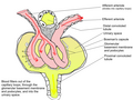
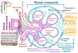 The structure of the renal corpuscle. | |
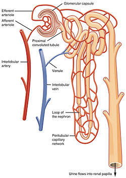 Diagram of the circulation related to a unmarried glomerulus, associated tubule, and collecting system The renal corpuscle in the cortex (outer layer) of the kidney. At the top, the renal corpuscle containing the glomerulus. The filtered blood exits into the renal tubule as filtrate, at correct. At left, blood flows from the afferent arteriole (red), enters into the renal corpuscle and is filtered in the glomerulus; blood flows out of the efferent arteriole (blue). | |
| Details | |
| Location | Nephron of the Kidney |
| Identifiers | |
| Latin | corpusculum renis |
| FMA | 15625 |
| Anatomical terminology [edit on Wikidata] | |
A renal corpuscle (also called malpighian trunk [1]) is the blood-filtering component of the nephron of the kidney. It consists of a glomerulus - a tuft of capillaries composed of endothelial cells, and a glomerular capsule known equally Bowman's capsule.[2]
Structure [edit]
The renal corpuscle is equanimous of two structures, the glomerulus and the Bowman'south sheathing.[3] The glomerulus is a pocket-size tuft of capillaries containing ii prison cell types. Endothelial cells, which have large fenestrae, are not covered by diaphragms. Mesangial cells are modified smooth muscle cells that lie between the capillaries. They regulate claret menstruum by their contractile activity and secrete extracellular matrix, prostaglandins, and cytokines. Mesangial cells also have phagocytic activity, removing proteins and other molecules trapped in the glomerular basement membrane or filtration barrier.
The Bowman's capsule has an outer parietal layer equanimous of uncomplicated squamous epithelium. The visceral layer, equanimous of modified unproblematic squamous epithelium, is lined by podocytes. Podocytes take foot processes, pedicels, that wrap effectually glomerular capillaries. These pedicels interdigitate with pedicels of side by side podocytes forming filtration slits.
At that place are two poles in the renal corpuscle, a vascular pole and a tubular pole. The vascular pole is a location of the glomerulus. At the vascular pole, the afferent arterioles and efferent arterioles enter and leave the glomerulus in the Bowman's capsule. The tubular pole is at the other end reverse to the vascular pole. At the tubular pole, the proximal convoluted tubule arises.[4]
Function [edit]
The renal corpuscle acts to filter blood. Fluid from blood in the glomerulus is nerveless in the Bowman'due south capsule to class "glomerular filtrate", which is and so farther candy along the nephron to form urine. It does this via a filtration bulwark. The renal corpuscle filtration barrier is composed of: the fenestrated endothelium of glomerular capillaries, the fused basal lamina of endothelial cells and podocytes, and the filtration slits of the podocytes. This barrier permits passage of water, ions, and small-scale molecules from the bloodstream into Bowman'southward space (the space between the visceral and parietal layers). Big and/or negatively charged proteins are prevented from passing into Bowman'due south space, thus retaining these proteins in the circulation. The basal lamina is composed of 3 layers: lamina rara externa, lamina densa, and lamina rara interna. The lamina rara externa is adjacent to the podocyte processes. The lamina densa is the primal layer consisting of blazon 4 collagen and laminin. This layer acts as a selective macromolecular filter, preventing the passage of big protein molecules into Bowman's space. The lamina rara interna is adjacent to endothelial cells. This layer contains heparan sulfate, a negatively charged glycosaminoglycan that contributes to the electrostatic barrier of the glomerular filter.
Eponym [edit]
A renal corpuscle is also known as a Malpighian corpuscle, named later Marcello Malpighi (1628–1694), an Italian physician and biologist. This proper name is no longer widely used, probably to avert confusion with a Malpighian corpuscle in the spleen.
Boosted images [edit]
-
Outline of function of renal corpuscle
References [edit]
- ^ "Renal corpuscle | anatomy". Encyclopedia Britannica . Retrieved 2021-04-15 .
- ^ J., Tortora, Gerard (2010). Principles of anatomy and physiology. Derrickson, Bryan. (twelfth ed.). Hoboken, NJ: John Wiley & Sons. p. 1024. ISBN9780470233474. OCLC 192027371.
- ^ "Renal corpuscle".
- ^ Mescher, Anthony L. (2016). Junqueira'southward Basic Histology, 14th edition. Lange. p. 397.
External links [edit]
- Histology image: 16003loa – Histology Learning System at Boston University
- MedEd at Loyola Histo/practical/kidney/hp16-53.html

Where Are Renal Corpuscles Found,
Source: https://en.wikipedia.org/wiki/Renal_corpuscle
Posted by: alexanderdellittef1972.blogspot.com
0 Response to "Where Are Renal Corpuscles Found"
Post a Comment